Near Field Optical Microscope Applications
Nsom Discovering New Worlds Test Measurement Photonics Handbook Photonics Buyers Guide
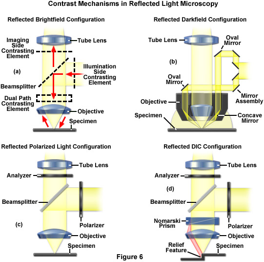
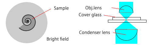
Zeiss Microscopy Online Campus Microscopy Basics Reflected Light Microscopy

Cell Biology Beyond The Diffraction Limit Near Field Scanning Optical Microscopy Journal Of Cell Science
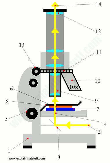
How Does A Microscope Work Explain That Stuff

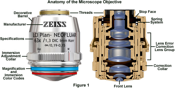
Lensless Microscopy Chip For Diagnostic Applications Medgadget In 2020 Microscopy Medical Tech Microscope Objective

Nano Antennas Assist In Improving Spatial Resolution Of Terahertz Microscopy Quantum Mechanics Nanotechnology Spatial

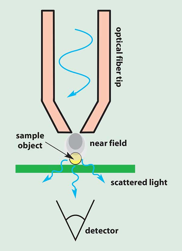
Aperture near field scanning optical microscopy nsom with fluorescence detection gives bio chemical specificity and orientational information in addition to the simultaneously acquired force image.
Near field optical microscope applications.

World S Most Powerful Optical Microscope Lets Researchers See Inside Viruses Popular Science

Nanotech Metasurfaces Will Usher In New Optic Technologies Technology Optical Microscope Physics Research
Zeiss Education In Microscopy And Digital Imaging

Since The Early 1930s Electron Microscopy Has Provided Unprecedented Access To The Alien Wo In 2020 Artificial Intelligence Algorithms Image Patch Systems Engineering
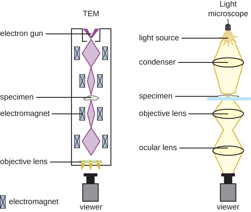
Zeiss Microscopy Online Campus Microscopy Basics Microscope Optical Systems
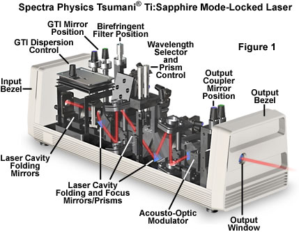
Molecular Expressions Microscopy Primer Specialized Microscopy Techniques Laser Systems For Optical Microscopy

Optical Microscopy An Overview Sciencedirect Topics
Limitations Of Optical Microscopy
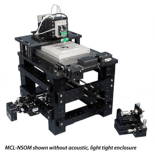
Mcl Nsom Near Field Scanning Optical Microscope

Microscopes An Overview Sciencedirect Topics

Nsom Snom History And Description Nanonics Imaging
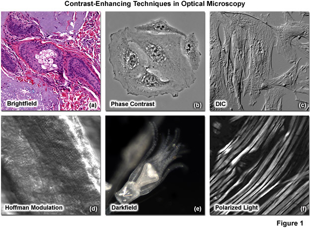
Zeiss Microscopy Online Campus Microscopy Basics Enhancing Contrast In Transmitted Light
Instruments Of Microscopy Microbiology

Seeing Atoms And Clusters On Surfaces Sciencedirect

Magnification Invariant Surface Profiling Technique For Structured Illumination Imaging And Microscopy Microscopy Magnification Biomedical Science

Understanding Microscopes And Objectives Edmund Optics
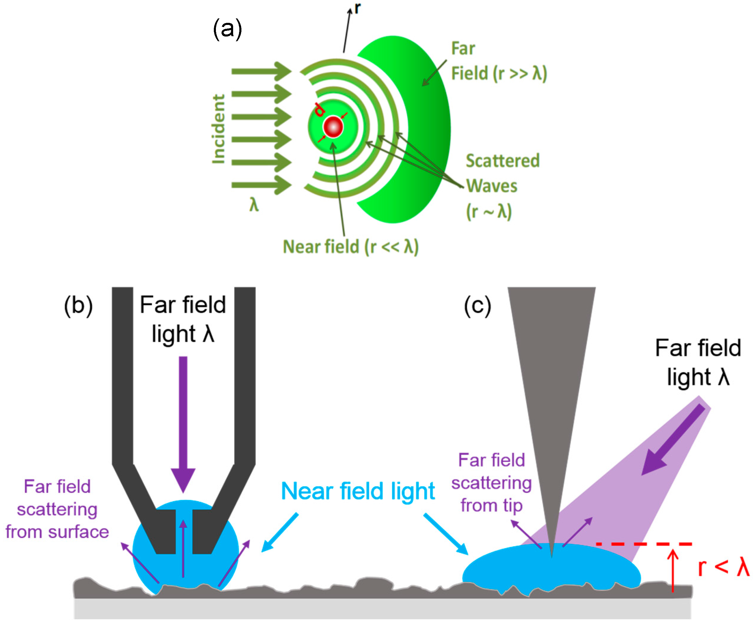
Applied Sciences Free Full Text A Review Of Three Dimensional Scanning Near Field Optical Microscopy 3d Snom And Its Applications In Nanoscale Light Management Html
Basic Of Optical Microscope Nanophoton Corp
Https Encrypted Tbn0 Gstatic Com Images Q Tbn 3aand9gct1kwsc Lmrakor3fa4eg7tm649b3jp1rc2ek81tmb8wjjz36j1 Usqp Cau

Angular Dispersions In Terahertz Metasurfaces Physics And Applications Advances In Engineering Physics Angular Scientific Articles

Quasi Monoenergetic And Tunable X Rays From A Laser Driven Compton Light Source X Ray Science Compton

Compound Microscope Definition Labeled Diagram Parts Uses

How To View Unstained Cells In Microscope Microscopic Annular Contrast
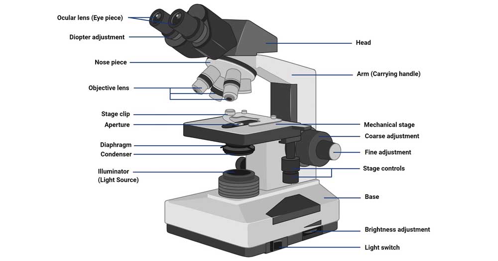
Parts Of A Microscope With Functions And Labeled Diagram
Source : pinterest.com
